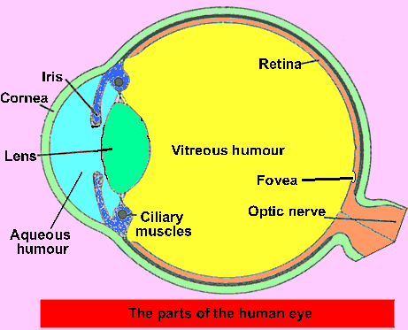
The lens acts to focus the image onto the retina (click to see an animation). It adjusts its shape so that close and far away objects are focused. When the lens ceases to function properly spectacles are prescribed.
The cornea is
a transparent part of the eye that along with the lens helps to focus
light on the retina.
The iris forms the colored part of the eye. Its function is to control the amount of light that travels through to the lens. The iris is composed of blood vessel, pigment and muscle fibres which control the size of the pupil (the black hole in the middle of the iris).
Cilliary muscles control the shape of the lens so that light is always focused on the retina.
The vitreous humour helps maintain the shape of the eye ball and helps keep the lens in position.
The aqueous humour helps maintain the shape of the cornea and to provide nutrients and oxygen to the cornea and lens that do not have blood vessels.
The retina is a light sensitive area at the back of the eye made of nerve cells. Two type of nerve cells predominate, rods and cones. Rods are sensitive to black and white while cones are sensitive to colour.
The fovea is the part of the retina that light is focused on. It consists mainly of nerve cells known as cones.
Why do you think
that the lens and cornea do not have blood vessels?
Why is it important
to control the size of the pupil?
Why do doctors recommend that you do not remove any object that has
penetrated the eye ball(such as a nail)?